Making Medicine Digestible

Category: Medicine

Auscultation of Heart Sounds : Methods, Areas, Interpretation, Differentiation

It is a dream of every medical student to one day be able to put a stethoscope on a patient and listen to their heart sounds, and this time correctly interpret it as well. In this article, we will go through the basics of how to auscultate (“Listening the sounds of internal organs from outside in an non-invasive manner”) heart sounds. In the upcoming posts, we will try to address the murmurs in detail. 

STETHOSCOPE :

Stethoscope is like an ornament for medical professionals and rightly so, It used to be one of the only tools to be able to diagnose complex medical conditions on the outpatient basis in a non-invasive manner. For a medical student of our generation, a stethoscope might not mean as much due to the invention of advanced diagnostics which are faster, far more accurate, objective in making decisions and becoming widely available. Still, auscultation is a primary care level practice widely used to screen patients for the presence of various conditions and in some cases, diagnose it. 

PARTS OF A STETHOSCOPE :

We will take a look at parts of the stethoscope for the starters. 

It mainly has following three parts :

1). Earpiece

2). Connecting PVC tubing

3). Chest piece (Has a larger part called Diaphragm and a smaller part called Bell)

Figure : Parts of a Stethoscope [Source : Britannica]

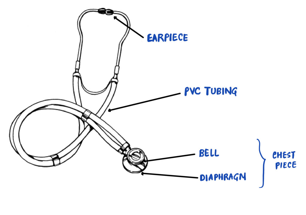
Chest Piece has two distinct parts which are meant to be used when detecting different pitched sounds. 

Low pitch sounds : Bell (Diaphragm can also detect low pitch sounds to a lesser degree)

High pitch sounds : Diaphragm 

HOW TO HOLD A STETHOSCOPE? :

Let’s first learn how to hold a stethoscope before starting discussion on auscultation findings.

Figure : Correct way to hold a stethoscope

Stethoscope is usually held with the neck connecting the diaphragm and the bell in between index and middle finger, as shown in the image below. Although it can vary as per Health Practitioner’s individual preference. 

Ear tips should be directed medially and forwards when in hand in order to minimise the tension in the ears. (You should not be able to see the holes in the tips when in the hand.)

Figure : Correct and incorrect method of placing ear tips [Source : Unikits]

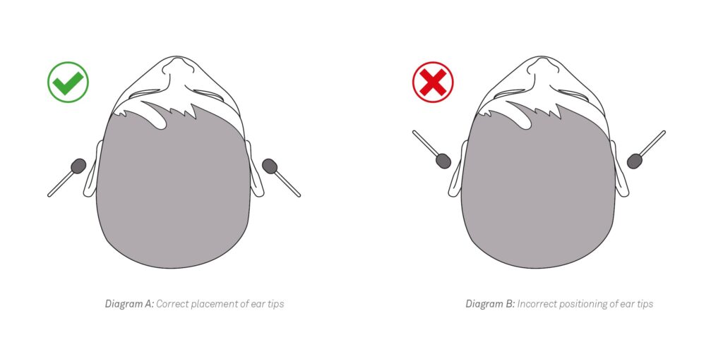
Care has to be taken not to press too much or too lightly while auscultation with a stethoscope. It just needs to be gentle enough so that you can appreciate the heart sounds. 


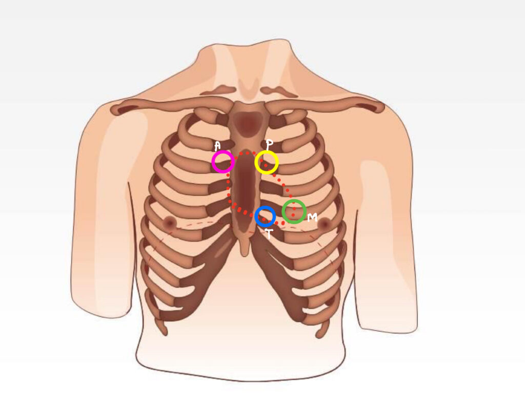
AREAS TO AUSCULTATE :

There are four areas to auscultate in a cardiac examination :

1). Mitral Area : Midclavicular line in 5th intercostal space (ICS) 

2). Tricuspid Area : Lower left sternal border, 5th ICS

3). Aortic Area : Upper right sternal border, 2nd ICS

4). Pulmonary Area : Upper left sternal border, 2md ICS

These areas reflect the position of the valve of the corresponding name. In theory, they correspond to the area of murmur of the respective valve pathology. However in practice, the distinction between these areas is not that distinct. For eg., Aortic valve regurgitation produces high pitched murmur that is audible from any of the above areas.

Figure : Figure showing different areas of auscultation including mitral (green), tricuspid (blue), aortic (pink) and pulmonary (yellow) area

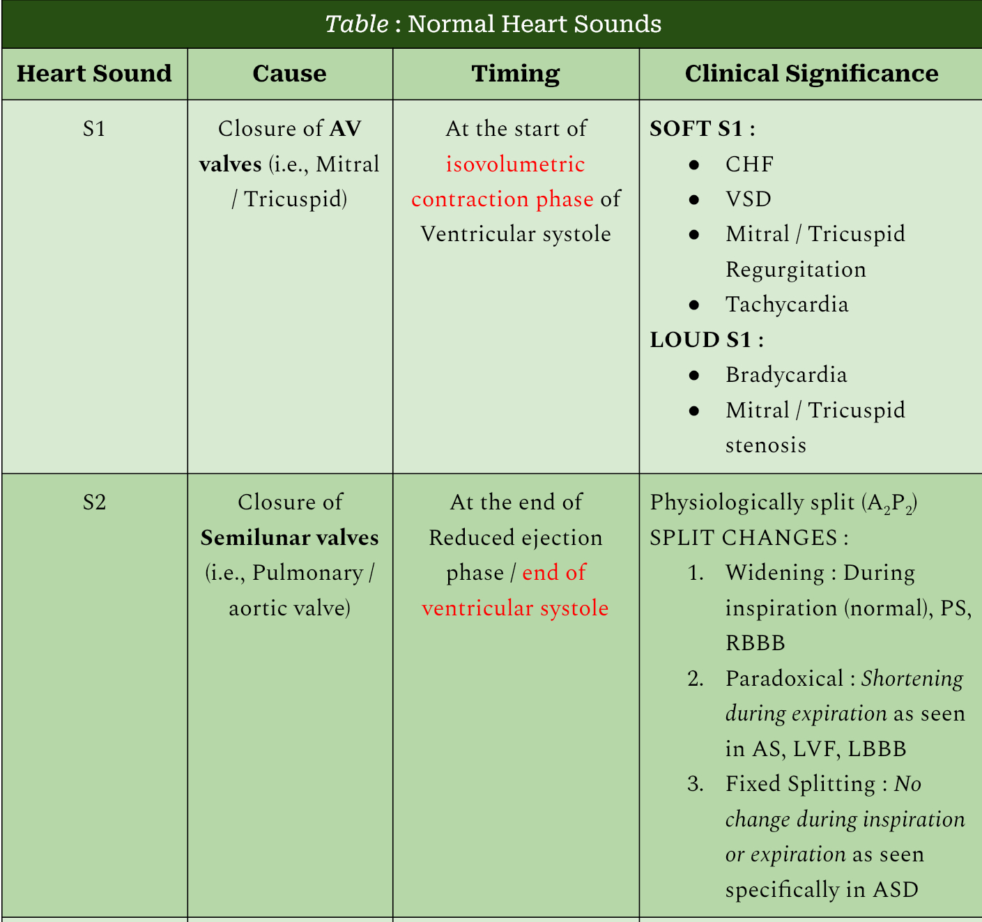
AUSCULTATING NORMAL HEART SOUNDS : 

First of all, it should be made clear that by “normal”, i mean the sounds that occur during the normal cardiac cycle and no other new sounds. These “normal” sounds can very well be representative of pathology as well (eg., loud S1 or Widely split S2). I have discussed these abnormalities in the post for Heart Sounds posted earlier. 

Table : Features of normal heart sounds S1 and S2 on auscultation

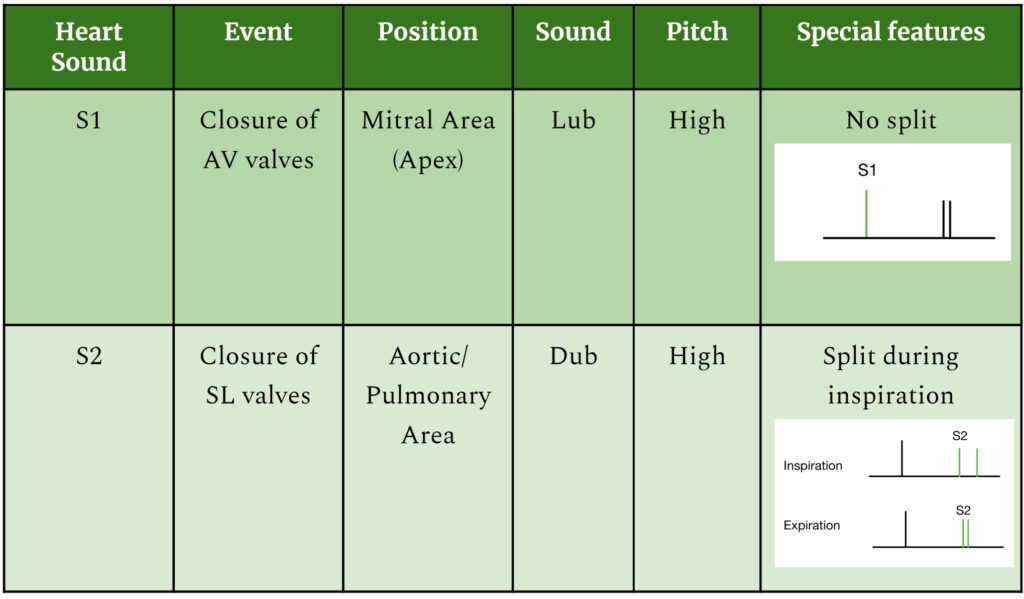
S1 and S2 can be easily recognized by their characteristic sounds “Lub…dub…lub…dub…” as in the audio given below.

Audio : Normal heart sound with clear S2 split

If you cannot understand what these sounds mean then there are other ways to differentiate these two :

  • Compare the pause in between the two sounds. Pause in between lub and dub is usually slightly shorter than the pause between dub and the next lub. Hence the correct way to identify it would be “lub..dub…….lub..dub…….lub..dub”, like that. 
  • S1 is louder than S2 that follows it.
  • You can ask the patient to take deep breaths in order to listen to the split of S2 clearly.

It will require practice in order to differentiate these two hence one last and definite way to learn to distinguish them is to just LISTEN MORE!!


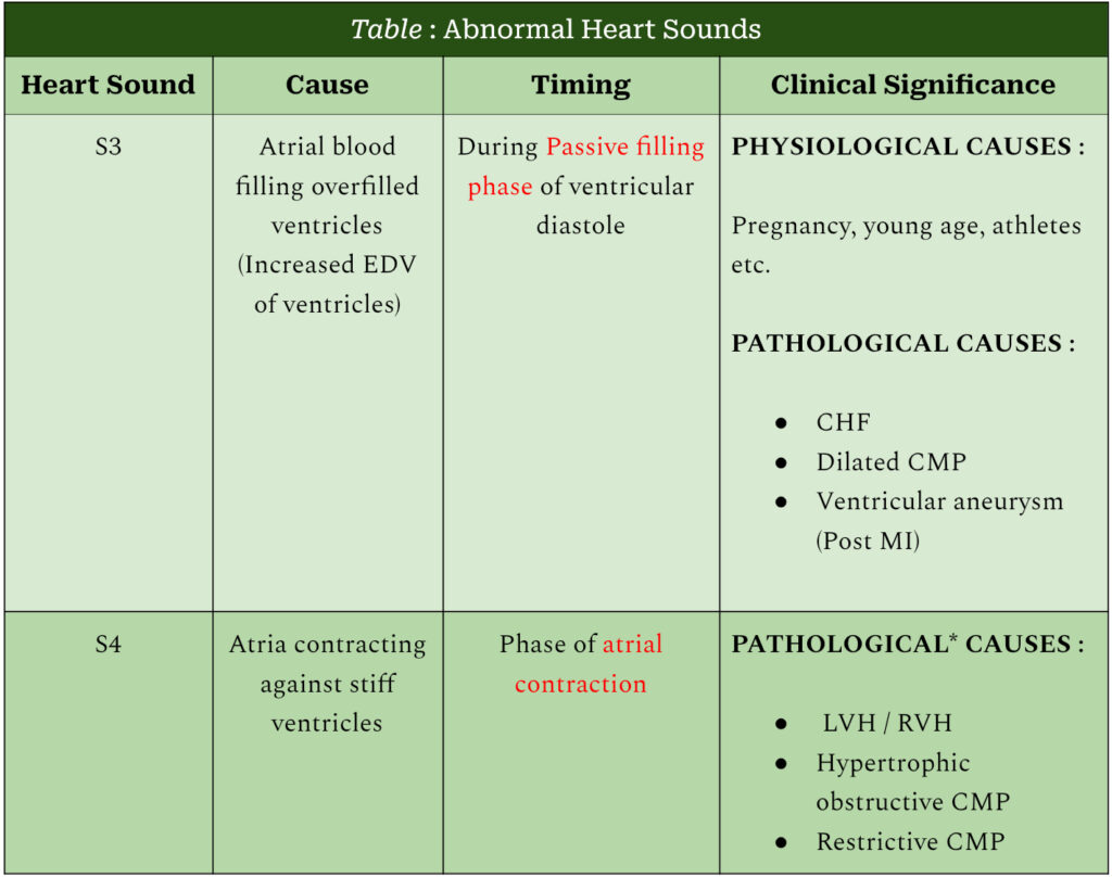
AUSCULTATING ABNORMAL HEART SOUNDS :

Apart from the above two sounds, a patient can have abnormal heart sounds which are result of some pathology or can be physiological as well. Details of the conditions that cause these sounds are discussed in the post covering Heart Sounds.

Table : Auscultatory features of S3 and S4 heart sounds

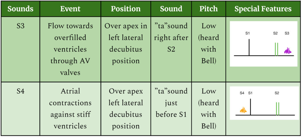
S3 when heard over apical area with the bell in left decubitus position goes like “lub…dub.ta……lub…dub.ta……lub…dub.ta”, like that.

Audio : S3 in LLD with Bell heard on Apex

S4 when heard in similar fashion, will be heard as “ta.lub…dub…..ta.lub…dub……ta.lub…dub”.

A genuine query arises while listening to S3 which is, “How to differentiate it from very similar sounding split S2, especially in cases of wide S2 split?”

It can be addressed in two different ways :

1). Practically it is not difficult to differentiate these two as their listening areas and parts of the stethoscope from which they are heard is completely different. 

S3 : Left lateral decubitus position in apical area with bell 

S2 split : Aortic / pulmonary area with diaphragm 

2). In case an audio clip is given in the exam, then one would have to rely largely on the history and rest of the physical examination given such as co-morbidities, signs of heart failure, whether the audio is recorded while performing deep inspiration and expirations or not etc. 

This sums up everything that you need to know in order to get started in auscultation of normal and abnormal heart sounds. Off course it is important to know the causes of abnormal heart sounds which are discussed in the other post by us. Do check that out before heading away.


SUMMARY AND STEPS OF EXAMINATION :

So once you are ready with your expensive stethoscope, take following steps in order to perform cardiac auscultation :

1). Check the functioning of the stethoscope. 

2). Explain the procedure to the patient with pointing out the areas where you are going to auscultate.

3). Keep patient in sitting position at the start.

4). Listen these areas in order : Mitral – Tricuspid – Aortic – Pulmonary

(Mnemonic : Multiple Types Are Present)

5). S1 and a soft S2 are heard at apical(mitral) position. Denote any change in the intensity of S1. Rule out the causes accordingly. 

6). S2 split is more appreciable in aortic / pulmonary areas and during inspiration. Note any changes in the normal split of S2. Rule out the causes accordingly. 

7). Move the patient to the left lateral decubitus position and note any abnormal heart sounds in the apical area. If present, rule out/in the causes accordingly. 


Please share your reviews on the article and suggestions if any. Do share it with your peers if you liked the explanation. Tell us how we can improve in the comments. See ya later!

Gas Gangrene : Etiology, Clinical Features, Diagnosis and Treatment

DEFINITION :

    Rapidly spreading, edematous myonecrosis, occurring in association with severely crushed wound contaminated with pathogenic clostridia.

OTHER NAMES :

Malignant edema / Clostridial Myonecrosis.

ETIOLOGY :

Gas gangrene is caused by clostridia species. Organisms involved can be typed in following manner :

Figure : Etiological agents involved in causation of Gas Gangrene

PATHOGENESIS :

Gas Gangrene usually follows a traumatic event. The steps that unfold during the pathogenesis of the disease are as follows :

Figure : Pathogenesis of Gas Gangrene and role of Alpha and Theta Toxins

CLINICAL MANIFESTATIONS :

Incubation Periods (period between contact and clinical manifestations) :

10-14 hrs (C. perfringens)

2-3 days (C. septicum)

5-6 days (C. novyi)

Various manifestations :

  • Sudden, excruciating pain
  • Foul-smelling serosanguineous discharge
  • Crepitus in muscle planes
  • Brawny edema and induration
  • Septic Shock

LABORATORY DIAGNOSIS :

It has only confirmatory and species identification roles. Treatment should be started immediately based on the clinical diagnosis.

Specimen :

Ideally from necrotic tissues, muscle fragments and exudates from deeper part of the wound

Blood culture may be positive for C. perfringens and C. septicum

Should be put into Robertson’s cooked meat broth and transferred immediately to the laboratory

Direct Microscopy :

  • Absence of neutrophils in the infected tissues is characteristic.
  • Thick, stubby, boxcar-shaped, gram-positive bacilli without spore- suggestive of C. perfringens are seen
  • Spore bearing gram-positive bacilli suggest other species :
  • Citron bodies– suggestive of C. septicum
  • Oval sub-terminal spores– suggest C. novyi

Identification for C. perfringens :

A. Target hemolysis (double zone hemolysis) :

Inner zone: due to theta-toxin

Outer zone: due to alpha-toxin

B. Nagler’s reaction – Opalescence surrounding streak line om egg yolk agar due to lecithinase activity of alpha-toxin

C. Reverse CAMP test – Arrow-shaped hemolysis pointing towards C. perfringens when Streptococcus agalactiae is streaked perpendicular to it.

D. Heat intolerance – C. perfringens grow when RCM broth is incubated at 45 degree Celsius for 4-6 hrs.

E. Stormy clot reaction in litmus milk due to lactose fermentation

TREATMENT :

  • Early surgical debridement (most crucial step)- Wound closure delayed until site is infection-free.
  • Penicillin with Clindamycin for 10-14 days.
  • Hyperbaric Oxygen against obligate anaerobes like C. perfringens but not on aerotolerant clostridia like C. septicum.
  • Anti alpha-toxin antiserum (Passive immunization).

The thoughtless person playing with penicillin treatment is morally responsible for the man who succumbs to death due to penicillin resistant organism.

Alexander flaming

REFERENCE :

  1. Apurba S Sastry, Sandhya Bhat, Essentials of Medical Microbiology, third edition, 2021 Jaypee Brothers Medical Publishers.

Heart sounds : Production, Timing and Clinical Significance

Heart sounds are simply the sounds that are generated during cardiac cycle by valvular events and flow of blood across it.”

IMPORTANCE OF HEART SOUNDS :

As heart sounds tell us about the normal functioning of the heart without any invasive procedure, they can be helpful to diagnose various clinical conditions of the heart on an office basis.

Before we had radiological diagnostic modalities, they used to be the sole source of diagnosis. 

One more important use of auscultation of heart sounds is to assess the severity of given valvular disease (eg., Aortic and pulmonary stenosis).

TYPES OF HEART SOUNDS AND THEIR PRODUCTION: 

S1 and S2 are the two main types of heart sounds (so called “normal / regular heart sounds”). The details of the normal heart sounds is given in aconcise manner in the below given table :

Table : Production, timing and clinical significance of normal Heart sounds (S1 & S2)

Apart from these, there can be other heart sounds which are usually an indicator of underlying pathology but can be normal as well. They are described in table below :

Table : Abnormal Heart sounds S3 % S4 (*Soft S4 might be physiologically present in some older individuals)

ADDITIONAL HEART SOUNDS : 

Apart from above mentioned basic types of heart sounds, there are some additional ones that can be heard during auscultation sometimes and they are enumerated with description as below :

A. EJECTION CLICK : 

Sound made by rapid flow of blood across the valve during rapid ejection phase.

It usually occurs in semilunar valves as ventricles have high pressures compared to atria. They are heard when end diastolic pressures in ventricles are high such as, increase in afterload (Aortic / Pulmonary stenosis). 

B. OPENING SNAP :

It’s atrial version of ejection click.

When atrial pressures are high, mitral / tricuspid valves open with a snap which is called opening snap (OS).

Conditions where OS might be present are mitral / tricuspid stenosis. It is heard during the early diastole phase of the cardiac cycle. 

C. MID-SYSTOLIC CLICK (NON-EJECTION CLICK) :

It is seen when ventricles have become sufficiently empty during the ejection phase and is specific for Mitral Valve Prolapse.

It has to be differentiated from ejection click, which is early in systole. 

TRACING OF VARIOUS HEART SOUNDS DURING CARDIAC CYCLE :

Below is a diagram that gives an idea regarding timing of various heart sounds, normal and abnormal, during cardiac cycle :

Figure : Diagrammatic tracing of different types of heart sounds according to their timing in cardiac cycle

IVC : Isovolumetric Contraction, EP : Ejection phase [Ventricular Systole]

IVR : Isovolumetric Relaxation, PFP : Passive Filling Phase, AC : Atrial Contraction [Ventricular Diastole]

EC : Ejection Click, MC : Mid-systolic Click, OS : Opening Snap,

With this, we finish the basics of heart sounds with their clinical significance and tracing on cardiac cycle.  

It is infinitely better to transplant a heart than to bury it to be devoured by worms.

Christian barnard

REFERENCES :

  1. Hall, John E. Guyton and Hall Textbook of Medical Physiology. 13th ed., Elsevier, 2016.
  2. Le, Tao; Bhushan, Vikas; and Sochat, Matthew. First Aid for the USMLE Step 1 2021. New York: McGraw-Hill Education, 2021.

Glycogen Storage Disorders : Types, Etiology, Clinical Features, Diagnosis and Management

Glycogen storage Disorders are biochemical disorders characterised by abnormal glycogen metabolism which can be presented by two major organs i.e, Liver and Muscle. In this particular section, we will provide a concise note on types of liver glycogen storage disorders aka Liver Glycogenosis.

Figure : Types of Glycogen Storage Disorders

LIVER GLYCOGENOSIS :

Figure : Types of Liver Glycogenosis with their respective enzyme deficiency / defects

TYPE 1 GSD : VON GIERKE’S DISEASE

INHERITANCE : Autosomal recessive

DEFICIENT ENZYME :

In subtype 1a, Glucose-6-phosphatase

In subtype 1b, Translocase (carries glucose-6-phosphate across microsomal membrane)

ORGANS AFFECTED : Liver, Kidney, Intestinal Mucosa

CLINICAL FINDINGS :

  1. Neonatal period: Hypoglycemia and Lactic Acidosis
  2. 3-4 months: Hepatomegaly
  3. Physical features: Doll-like facies, fat cheeks, thin extremities, short stature, protuberant abdomen
  4. Growth retardation
  5. Easy bruising and epistaxis
  6. Diarrhoea and malnutrition (long-standing cases of subtype 1b)

LABORATORY FINDINGS :

  1. Prolonged bleeding time
  2. Hyperuricemia
  3. Elevation of TGs, LDL, Total cholesterol
  4. In type 1b, neutropenia (leading to recurrent bacterial infections and mucosal ulcerations) 

LONG TERM COMPLICATIONS :

  1. Gout (presented at puberty)
  2. Polycystic ovaries and menorrhagia in women
  3. Pancreatitis
  4. Systemic hypertension
  5. Osteopenia/osteoporosis (seen in adults)
  6. Hepatic adenomas (2nd-3rd decade of life)
  7. End stage renal disease in patients who developed proteinuria, nephrocalcinosis and alteration in creatinine clearance

DIAGNOSIS :

  1. Clinical presentation
  2. Lab findings (mentioned above)
  3. Genetic testing
  4. Liver biopsy (historic value)- Distended by glycogen and fat, with large lipid vacuoles

TREATMENT :

  1. First line: Avoid fasting and frequent feedings
  2. Diet preferred:  Complex carbohydrates with uncooked corn-starch
  3. For nephrocalcinosis: Citrate supplementation
  4. For hyperuricemia: Allopurinol
  5. For lipid abnormalities: HMG-CoA reductase inhibitors and fibrates
  6. For microalbuminuria: ACE inhibitors
  7. For liver adenoma: Surgical resection, radiofrequency ablation, percutaneous ethanol injection
  8. Liver and Kidney transplantation may be indicated
  9. For subtype 1b: G-CSF and Empagliflozin 

TYPE 3 GSD : CORI’S DISEASE

INHERITANCE : Autosomal recessive

ENZYME INVOLVED : Glycogen debranching enzyme

CLASSIFICATION :

3a: Liver, skeletal muscle and cardiac involvement

3b: Primarily liver involvement

CLINICAL FINDINGS :

  1. Infancy: Hypoglycemia(ketotic or non-ketotic in only 50% of patients) , hepatomegaly (reduces with age), hyperlipidemia, short stature
  2. 3a: Skeletal myopathy and cardiomyopathy may be present with the above findings
  3. Liver: Hepatomegaly reduces with age but many patients in late adulthood have fibrosis, cirrhosis, liver failure, hepatocellular carcinoma, hepatic adenomas( less common than in Von Gierke’s disease)
  4. Heart: Left ventricular hypertrophy, arrhythmia
  5. Skeletomuscular: Muscle weakness (severe by 4th decade), exercise intolerance, osteoporosis
  6. Nervous system: Peripheral Neuropathy
  7. Polycystic ovaries in females

LABORATORY FINDINGS :

  1. Elevated ALT and AST
  2. Normal blood lactate and uric acid
  3. Elevation of TGs, LDL, Total cholesterol
  4. Serum creatine kinase may be elevated

DIAGNOSIS :

  1. Clinical presentation
  2. Lab findings (mentioned above)
  3. Genetic testing- DNA based analyses
  4. Liver biopsy (historic value)- distended hepatocytes, periportal fibrosis along with few fat infiltration 

TREATMENT :

  1. Diet preferred:  As gluconeogenesis is intact, high-protein diet and complex carbohydrates with uncooked corn-starch
  2. Dietary lipid modifications: High fat diet/ketogenic diet/supplementation of medium-chain TGs
  3. For lipid abnormalities: HMG-CoA reductase inhibitors and fibrates
  4. For liver adenoma: Surgical resection, radiofrequency ablation, percutaneous ethanol injection
  5. Liver and heart transplantation may be indicated

TYPE 4 GSD : ANDERSON’S DISEASE

ENZYME DEFICIENT : Branching Enzymes

CLASSIFICATION WITH FEATURES :

  1. Hepatic form- Failure to thrive, hypotonia, hepatomegaly, cirrhosis, failure (Death by 5 years), hypoglycemia (late finding secondary to liver lesions); small subset has extrahepatic involvement
  1. Neuromuscular forms-

It has 4 subtypes: 

  1. Perinatal– Death in neonatal period
  2. Congenital– Death in neonatal period
  3. Childhood– Myopathy, cardiomyopathy, systemic findings
  4. Adult Polyglucosan Body Disease (APBD)– B/L Lower limb weakness, spasticity; neurogenic bladder; peripheral neuropathy; cognitive impairment

DEFINITIVE DIAGNOSIS :

  1. Demonstrate pathogenic variants in the GBE1 gene; or
  2. Enzyme deficiency in liver, muscle, cultured skin fibroblasts, leukocytes

TREATMENT :

  1. Liver transplantation
  2. Symptomatic for gait abnormalities and bladder dysfunction in APBD 

TYPE 9 GSD

ENZYME INVOLVED : Liver Phosphorylase Kinase (PhK)

CLASSIFICATION :

  1. 9 alpha 2 (PHKA2 pathogenic variants): 
  • X-linked 
  • commonly seen
  • high phenotypic variability
  • Enzymes in liver, erythrocyte, leukocyte affected, not muscles
  1. 9 beta (PHKB pathogenic variants)
  2. 9 gamma 2 (PHKG2 pathogenic variants): Severe with early cirrhosis and fibrosis
  3. 9 alpha 1 (PRKAG2 pathogenic variants): Affect only muscles
  4. 9 gamma 1 (PRKAG2 pathogenic variants): Affect only muscles

TYPE 10 GSD : FANCONI-BICKEL SYNDROME

ENZYMES DEFICIENT : GLUT2 deficiency

FEATURES : Proximal renal tubular dysfunction leading to increased renal clearance of glucose, amino acids, phosphate and uric acid

Medicine is a science of uncertainty and an art of probability.

William osler

REFERENCE :

Harrison’s Principles of Internal Medicine, 21e Loscalzo J, Fauci A, Kasper D, Hauser S, Longo D, Jameson J. Loscalzo J, & Fauci A, & Kasper D, & Hauser S, & Longo D, & Jameson J(Eds.),Eds. Joseph Loscalzo, et al.

An easy way to remember the locations of lymphocytes within the Lymph node

Lymphocytes occupy specific regions within the lymph node and it can be quite tiring at times to remember which one lies where. A small mnemonic can be used in order to keep the information tidy in your brain castle.

Figure : Diagram of lymph node showing locations of B cells and T cells within follicles and paracortex, respectively

The mnemonic goes : Boy Friend (needs) Toilet Paper

B cells reside in Follicles of the lymph nodes which are also known as primary follicles. They form the definitive Cortex of the lymph node. They also occupy the Medulla (the center most part) of the lymph node.

The area in between cortex and paracortex is the home for T cells of the lymph node, called Paracortex.

Powered by WordPress & Theme by Anders Norén